
![]()
Gutener is clean, creative, powerful, flexible, highly customizable and Gutenberg ready WordPress theme. It is fully responsive and is sure to make your website stand out from the crowd.
Common Retinal Issues
The most common type of retinal disorders are Macular degeneration, Diabetic Retinopathy, Retinal Scars, etc.
RKEC Advantage
At RK Eye Centre, we have embraced the rapid strides in technology especially in the field of diagnostic imaging of the retina and vitreo retinal surgery. We have the capacity to take pictures of the back of the eyes and even take optical slices of the retina without any direct contact. Even if the retina cannot be visualised due to various reasons like corneal scars, or cataracts, we have the capability of getting high resolution scanned images of the entire eye
Diagnostic Technology
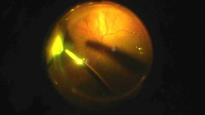
1. Fundus Photography – Kowa, Japan
Permits us to procure color photographs and red free images of the back of the eye.
2. Fluorescein Angiography (FFA) – Kowa, Japan
This diagnostic procedure enables us to document inconsistencies in the blood circulation of the retina. It is very similar to a coronary angiogram for checking blood circulation to the heart. It involves the injection of a dye called Fluorescein into a vein of the arm following which repeated photos of the retina are taken. It is very useful in the management of diabetic retinopathy.
3. High definition Optical Coherence Tomography (HD-OCT) – Zeiss, USA
We have come a long way in understanding what the retina looks like. Imagine being able to take a slice of the back of the eye without even touching it. The Zeiss, Optical Coherence Tomography uses a beam of light and its reflection to obtain cross-sectional images which provide information about the different layers of the retina.
4. B-scan Ultrasonography – Quantel, France
This procedure involves use of high-frequency sound waves to examine the eye when normal view is obscured by hemorrhage or cataracts.
Surgical Technology
Laser Procedures
With Gold standard single spot delivery system we have the technology to treat many retinal diseases like diabetic retinopathy, retinal holes and tears, without the need of performing invasive surgery. The addition of an Indirect Ophthalmoscope delivery system enables us to reach even the toughest corners of the retina. Most of these are performed under topical anesthesia (drops) and as outpatient procedures.
Intravenal Injections
A lot has changed in the area of intra vitreal injections and implants. These have come a long way in the management of conditions like age related macular degeneration, diabetic macular edema and vascular occlusions in the eye. We routinely inject Lucentis (Ranibizumab), Avastin (Bevacizumab), Macugen (Pegaptinib Sodium), Ozurdex (steroid implant) and Triamcinolone acetonide (Depot steroid).
Surgical Procedures
A wide range of retinal procedures are performed with the Alcon Constellation system which is the top of the line vitrectomy system. We possess the capability to perform sutureless vitrectomy even in the most complex of cases. We can now perform procedures like:
Complex retinal detachment surgery
Diabetic vitrectomies
Dislocated nucleus and intraocular lenses retrieval
Scleral buckle and scleral fixated lenses
Vitreous Issues
Vitreous Liquification
Most common degenerative change in the vitreous. Appearance of coarse aggregate material which moves freely in the vitreous.
Causes: Myopic degeneration, Retinotis pigmentosa.
Post inflammatory condition following ureitis.
Trauma or Radiation
Posterior Vitreous degeneration
Separation of the vitreous from the back of the eye. Vitreous shrinks causing development of cloudy clumps in vitreous.
Symptoms: Floaters and flashes
Vitreous Opacities
Any non transparent structure present in the vitreous it will form an opacity due to its transparent nature.
1. Inflammatory vitreous opacities: Exudates paired into the vitreous in anterior ureitis and posterior ureitis
2. Amyloid degeneration: Amorphous amyloid material is deposited in the vitreous.
3. Asteroid hyalosis: It is some small rounded bodies suspended in the vitreous gel. It is formed due to accumulation of calcium containing lipid.
Besides we have a full time vitreo retinal specialist with us to take care of your retinal needs.
Diabetes causes blood vessels in the retina to leak. The leaky blood vessels in the retina associated with diabetes is termed diabetic retinopathy
Most diabetics have some form of diabetic retinopathy. It may vary from mild to severe. It tends to develop about 7 – 10 years from the diagnosis of diabetes.
It can be controlled to a certain degree by effective control of sugar levels. The progression of diabetic retinopathy can be stalled through effective control and can rarely be prevented as well. The effect of diabetic control and its relationship to diabetic retinopathy varies from individual to individual and is probably linked to gene composition.
It is categorised into non proliferative diabetic retinopathy (NPDR) and proliferative diabetic retinopathy (PDR). There are 4 stages of non proliferative diabetic retinopathy ranging from mild to very severe. Proliferative diabetic retinopathy can be classified as early or high risk depending on the extent of new blood vessel formation. Advanced diabetic eye disease (ADED) is end stage of diabetic retinopathy leading to blindness.
Strict control of blood glucose levels is the most important in the management of diabetic retinopathy. Other treatment options include laser treatment, injection of intravitreal drugs and vitreo retinal surgery.
Laser is generally indicated when there are leaking microaneurysms close to the fovea (most important point on the retina where most light is focused) or if there are presence of new blood vessels on the retina.
When there are elements of traction involved or membrane formation over the retina or if there is profuse bleeding inside the eye, then surgery is usually required.
You wouldn’t. The symptoms of retinopathy would arise only when crucial parts of the retina (the fovea) are involved. SInce retina is a relatively large structure, it may take months for existing damage in non critical areas to spread to the fovea. Also some may have blackish or reddish spots disturbing vision. this may indicate the presence of blood in the vitreous requiring urgent attention

At RK Eye Centre, we have always strived to keep up with the advancements in the
field of cataract surgery….

With cosmetic correction of refractive error becoming more and more acceptable among the general public we offer .

Glaucoma is a group of eye diseases that is eventually cause damage to the optic
nerve. Most commonly it is caused..
At RK Eye Centre, we have embraced the rapid strides in technology especially in the field of diagnostic…

With an increase in the demand for cosmetic surgery we offer a range of procedures
like..

This super speciality offers the latest in the management of various eye problems seen
in the Pediatric..

Various corneal problems like infectious keratitis (infections of the cornea), Autoimmune
related peripheral..

This super speciality field deals with the nerve behind the eye and the relationship
between the eye and the brain.