LASIK is a well-known laser refractive surgery to improve the vision of the patient and to correct the problem of refractive error. LASIK is typically performed to rectify myopia, hyperopia, and astigmatism. A perfect candidate can expect good long-term results from the surgery and to get the best results various stages of evaluation are performed to select a good candidate. The preoperative evaluation is well structured and is an important part of the LASIK surgery process itself. Majorly the evaluation can be classified into different stages – a History of the patient, Physical examination, and Pre LASIK testing.
History of the patient:
It is vital to have knowledge of the patient’s expectations/results of the LASIK surgery,
Past medical history of the patient:
Past medical information is necessary prior to any surgery to better understand the outcomes of the procedure. Some systemic diseases like autoimmune diseases, collagen vascular diseases, and diabetes could potentially affect the healing process, due to the disturbance the results may be prolonged.
On the other hand, some diseases can reoccur after surgery. For instance, peripheral keratitis and ocular herpes may recur after the LASIK. The patient must be made aware of these risks.
Information about their medications:
Intake of several drugs could influence the results of the surgery and to avoid this risk, a complete list of the patient’s medications is noted.
Social and family history:
Enquiries with regards to the lifestyle of the patient are made to ascertain the excessive use of alcohol and cigarettes as this could interrupt the healing process of the surgery.
To avoid the risk of flap complications, information about high-risk activities like contact sports are collected. Some familial diseases may be present and expressed in varying degrees of a family and the presence of such diseases could affect the outcomes of LASIK surgery.
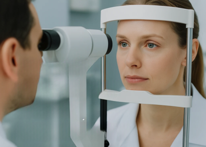
Physical examination:
Visual acuity: Uncorrected (without correcting refractive index) and corrected (correcting the refractive index) are taken.
Refraction test: A clear refraction check is completed first and a cycloplegic refraction check is done later if required.
Complete eye examination: Every patient undergoes an absolute, thorough eye examination. The attributes of all eye-related structures from the eyelids to the retina are checked thoroughly. If any disease or infection is diagnosed, the particular problem should be treated promptly. The LASIK surgery can not be operated on if the patient suffers from a cataract, in such cases, the patient will be advised of other alternative methods of treatment.
LASIK testing:
Test for dry eyes:
The quality of the tear film is measured by the tear break-up time, Schirmer’s test, and MGD assessment. If any abnormalities are seen, then treatment in the form of lid hygiene and antibiotics is begun. The patients will be re-examined several times until the improvement of dryness.
Contrast sensitivity test:
The particular test is conducted to have a keen knowledge about the functions of the vision of the patient. The two major tests performed today are the Hamilton-Veale contrast sensitivity test and the Pelli Robson Chart.
Pupil test:
An infrared pupillometer is a device used to measure the size of the pupil accurately. Pupil size is tested because the large pupil size may lead to glare after the surgery. So it is important to know the size of the pupil to adjust the size of the optical zone of the laser.
Pachymeter:
An actual corneal thickness must be evaluated for the treatment, because the patients who have a thin cornea, have the risk of ectasia after the LASIK surgery.
Keratometry:
The shape of the cornea is manually evaluated as a preoperative process. The postoperative evaluation depends on the type and level of the LASIK treatment.
Wavefront analysis:
The test is conducted to determine the higher-order aberration, a patient with the notable higher order of aberration cannot be treated with standard LASIK procedures because they can end up with undesirable visual symptoms.
Corneal topography:
It is a three-dimensional imaging tool that is used to plot the surface of the patient’s cornea. It is used to detect a variety of corneal abnormalities. In some abnormalities like keratoconus, the LASIK surgery cannot be performed, in such cases, the LASIK may lead to the loss of vision.