Computers have increasingly become a part of our lives in the recent times. It is not possible to entirely avoid computers because of their significance they hold in our jobs and lives. 50 years ago, no one might have thought of getting health care while sitting in their house. But today that is precisely what you are doing at the moment. While it is an undeniable fact that computers have made a lot of jobs easy, we also need to realise the effects of their prolonged use on our health.
Seeing or working on computer for a long time may require a lot of work and strain from your muscles. Over the years, such strain may make your eye muscles less flexible. The brightness, the glares and the contrast do further damage to your eyes. Here are some easy tips to protect your eyes from such damage.
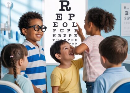
1. Getting your eyes examined regularly:
Having an annual eye test ensures that you detect eye problems, if any, at the earliest stage possible. If your job is one that involves a lot of computer usage, a regular check-up becomes even more important. Getting a comprehensive eye exam at least once a year is a necessity with this rapid digitalization. Measure how far your eyes are from your screen when you sit at your computer, and bring this measurement to your exam so your eye doctor can test your eyes at that specific working distance.
2. Proper lighting:
Avoid excessively bright lights at home and workplace. Try to get an anti-glare screen to minimise the effect of glare on your eyes. Avoid overhead lights while working on the system. Adjust the brightness and font size at an optimum level to avoid strain. If possible, position your computer monitor or screen so windows are to the side, instead of in front or behind it.
3. Blink more often:
When working at a computer, people blink less frequently – about one-third as often as they normally do. Blinking is very important when working at a computer; blinking moistens your eyes to prevent dryness and irritation. Tears coating the eye evaporate more rapidly during long non-blinking phases and this can cause dry eyes.To reduce your risk of dry eyes during computer use, try this exercise: Every 20 minutes, blink 10 times by closing your eyes as if falling asleep (very slowly). This will help lubricate your eyes.
4. Eye Exercise:
To reduce your risk of tiring your eyes by constantly focusing on your screen, look away from your computer at least every 20 minutes and gaze at a distant object (at least 20 feet away) for at least 20 seconds. Some eye doctors call this the “20-20-20 rule.” Looking far away relaxes the focusing muscle inside the eye to reduce fatigue.
Another exercise is to look far away at an object for 10-15 seconds, then gaze at something up close for 10-15 seconds. Then look back at the distant object. Do this 10 times. This exercise reduces the risk of your eyes’ focusing ability to “lock up” (a condition called accommodative spasm) after prolonged computer work.
5. Frequent breaks:
Many workers take only two 15-minute breaks from their computer throughout their work day. According to a recent study, discomfort and eye strain were significantly reduced when computer workers took four additional five-minute “mini-breaks” throughout their work day.
And these supplementary breaks did not reduce the workers’ productivity. Data entry speed was significantly faster as a result of the extra breaks, so work output was maintained even though the workers had 20 extra minutes of break time each day. During your computer breaks, stand up, move about and stretch your arms, legs, back, neck and shoulders to reduce tension and muscle fatigue.
6. Position:
Check your monitor’s position – It is important that your monitor be positioned at the proper distance away from your eyes. Optimally, your computer screen should be 15 to 20 degrees below eye level (about 4 or 5 inches) as measured from the center of the screen and 20 to 28 inches from the eyes. Improper posture during computer work also contributes to computer vision syndrome. Adjust your workstation and chair to the correct height.
Never postpone your doctor consultation once you feel there is something is wrong with your sight. Get your eyes tested at least once a year to detect any problems at the earliest stage possible. Get the best treatment from the best Eye Hospital in Chennai- RK Eye Centre. Our highly qualified team of doctors ensure that every patient gets the best possible treatment. RK Eye Centre was founded by Dr. Rajinikantha- one of the top 3 ophthalmologists in Chennai.
WHAT’S THE DEAL ABOUT GLAUCOMA?
Recently we get to hear the word ‘glaucoma’ often. What is this glaucoma? What are the symptoms of glaucoma? How is it diagnosed and how is it treated? This blog post shall answer all your queries. But this blog post’s intention doesn’t stop with you reading this. Get your eyes tested at a good eye hospital. If you are in Chennai the best choice you can make for your eyes is visiting R K Eye Centre. From patient treatment to the technical facilities, RK Eye Centre always gives the best to its patients.
What is Glaucoma?
An eye disease often associated with elevated intraocular pressure in which, damage to the optic nerve can lead to the loss of vision and even blindness. Intraocular pressure increases when either too much fluid is produced in the eye or the drainage or outflow channels of the eyes become blocked. It is the leading cause of irreversible blindness in the world. Some people can have optic nerves that are sensitive to normal eye pressure. This means their risk of getting glaucoma is higher than normal. Regular eye exams are important to find early signs of damage to their optic nerve.
Types of Glaucoma:
Primary open-angle glaucoma
This is the most common type of glaucoma. It happens gradually, where the eye does not drain fluid as well as it should (like a clogged drain). As a result, eye pressure builds and starts to damage the optic nerve. This type of glaucoma is painless and causes no vision changes at first.
Angle-closure glaucoma/ closed-angle glaucoma/ narrow-angle glaucoma
This type happens when someone’s iris is very close to the drainage angle in their eye. The iris can end up blocking the drainage angle. You can think of it like a piece of paper sliding over a sink drain. When the drainage angle gets completely blocked, eye pressure rises very quickly. This is an acute attack. In this case, you should consult your ophthalmologist immediately.
The signs of an acute angle-closure glaucoma attack:
- Sudden blurry vision
- Severe eye pain
- Headache
- Nausea or vomit
- Rainbow-colored rings or halos around lights
Many people with angle-closure glaucoma develop it slowly. This is called chronic angle-closure glaucoma. There are no symptoms at first, so they don’t know they have it until the damage is severe or they have an attack.
How common is glaucoma?
The number for those blinded by this disease stands at a staggering 6 million. Many of those diagnosed with this problem were not even aware of any difficulty with their vision. While everyone is at risk for glaucoma, certain people are at a much higher risk and need to be checked more frequently by their eye doctor. The major risk factors for glaucoma include the following:
- Age over 45 years
- Family history of glaucoma
- Diabetes
- History of elevated intraocular pressure
- Decrease in corneal thickness and rigidity
- Nearsightedness (high degree of myopia), the inability to see distant objects clearly
- History of injury to the eye
- Use of cortisone (steroids), either in the eye or systemically (orally or injected)
- Farsightedness (hyperopia), which is seeing distant objects better than close ones (Farsighted people may have narrow drainage angles, which predispose them to acute [sudden] attacks of angle-closure glaucoma.)
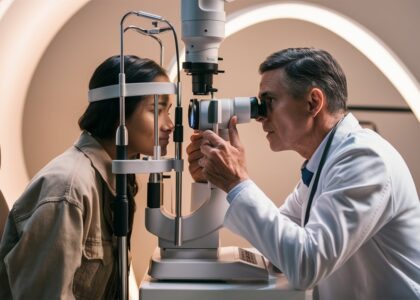
Diagnosis:
As most of the glaucoma cases present no symptoms, the only way to detect glaucoma is by getting a comprehensive eye exam done. A comprehensive glaucoma eye exam consists of the following tests:
- Tonometry (the inner eye pressure)
- Ophthalmoscopy / dilated eye exam (the shape and colour of optic nerve)
- Perimetry (the complete field of vision)
- Gonioscopy (the angle in the eye where the iris meets the cornea)
- Pachymetry (thickness of the cornea)
- Disc photography (documents current status of optic disc)
- Optical Coherence Tomography (detects early damage by directly measuring Retinal nerve fibre layer thickness)
Treatment:
The usual treatment for glaucoma is eye drops, although lasers and surgery can also be used. Most cases can be controlled well with these treatments, thereby preventing further loss of vision. A medicine for glaucoma is still being researched in various parts of the world. Early diagnosis is the only thing to catch glaucoma at the earliest stage possible and prevent it from progressing.
Now that you know better about glaucoma, don’t delay your eye exam anymore. To get the best experience and service with regards to eye problems including glaucoma, RK Eye Centre is the best place for people in Chennai. Get your eyes tested by the top eye specialists in the city, at RK Eye Centre.
EYE CARE FOR PEOPLE WITH DIABETES
When a normal person goes for his regular blood test, his first prayer is ‘God, please.. Not diabetes.. Please..!”
Diabetes is every person’s nightmare. For it doesn’t come alone. Diabetic people need to be careful about a lot of accompanying conditions. Diabetic people need to be concerned about a few eye problems in particular- diabetic retinopathy, diabetic macular edema, cataract and glaucoma. Lets see what these are.
Diabetic retinopathy is a problem wherein the retinal blood vessels leak fluid or bleed. Chronically high blood sugar from diabetes is associated with damage to the tiny blood vessels in the retina, leading to diabetic retinopathy. The retina detects light and converts it to signals sent through the optic nerve to the brain. Diabetic retinopathy can cause blood vessels in the retina to leak fluid or hemorrhage (bleed), distorting vision. In its most advanced stage, new abnormal blood vessels proliferate (increase in number) on the surface of the retina, which can lead to scarring and cell loss in the retina.In the advanced stage of diabetic retinopathy, the number of abnormal blood vessels or those leaking/ bleeding may increase, on the surface of the retina. This leads to scarring and cell loss in the retina. This disease is the most common cause of loss of vision among diabetic people.
Diabetic retinopathy usually gives no symptoms during the initial stages. Therefore it is advisable for diabetic people to get a comprehensive dilated eye exam at least once a year, so that it can be stopped at the earliest stage possible. Controlling diabetes can slow the onset and worsening of diabetic retinopathy.
Diabetic Macular Edema happens when diabetic retinopathy causes swelling in the macula area of the retina. It is the buildup of fluid (edema) in the macula. The macula is important for the sharp, straight-ahead vision that is used for reading, recognizing faces, and driving. About half of the people with diabetic retinopathy are likely to develop diabetic macular edema. Although in most of the cases it happens with worsening diabetic retinopathy, it is possible for diabetic macular edema to develop during any stage of the former problem.
Tractional Retinal Detachment: This refers to advanced diabetic eye disease when the gel like substance in front of the retina called the vitreous body becomes more fibrous like and pulls on the retina leading to its detachment. Such issues can be corrected only through Surgical procedures such as MIVS (Micro Incision Vitrectomy Surgery). The latest 25G and 27G (thinnest) vitrectomy procedures ensure faster healing time which is so crucial for a diabetic individual. At RK Eye Centre our Alcon, Constellation system US, top of the line system, provides the ideal platform for performing difficult vitrectomy procedures.
Diabetic retinopathy and Diabetic Macular Edema are detected during a comprehensive dilated eye exam that includes:
Visual acuity testing: This eye chart test measures a person’s ability to see at various distances.
Tonometry: This test measures pressure inside the eye.
Pupil dilation: Drops placed on the eye’s surface dilate (widen) the pupil, allowing a physician to examine the retina and optic nerve.
Optical coherence tomography (OCT): This technique is similar to ultrasound but uses light waves instead of sound waves to capture images of tissues inside the body. OCT provides detailed images of tissues that can be penetrated by light, such as the eye.
A comprehensive dilated eye exam allows the doctor to check the retina for:
- Changes to blood vessels
- Leaking blood vessels or warning signs of leaky blood vessels, such as fatty deposits
- Swelling of the macula (DME)
- Changes in the lens
Cataract is the clouding of the eye’s lens by debris. This leads to unclear vision and improper focus. Adults with diabetes are more likely to develop cataract than non-diabetic adults. But cataract is not necessarily limited to adults. Cataract may even develop in young people with diabetes.
Glaucoma is an eye problem that attacks the optic nerve. Some types of glaucoma may be caused by elevated blood pressure, but diabetes doubles the risk of glaucoma.
Gaining awareness of the possible problems is the first step towards preventing them. Never miss your annual check up, especially if you are diabetic. Semi annual check ups are recommended for patients with moderate disease process. A check up every 3 months is warranted for those with severe disease. Keep your blood sugar levels under control. Get yourself a comprehensive eye exam at RK Eye Centre– one of the best Eye Hospitals in Chennai. Our highly qualified and experienced doctors and trained staff will make sure you get the best quality of service possible. Once you come to RK Eye Centre, you no longer have the burden of worrying about you eye health. Leave it to us to give you the best treatment.
TIPS FOR HEALTHY EYES
1. Right Diet
Eating a diet rich in fruits and vegetables, particularly dark leafy greens such as spinach is important for keeping your eyes healthy. There are eye health benefits from eating fish high in omega-3 fatty acids, such as salmon, tuna, and halibut.
2. Wear protective eyewear
Wear protective eyewear when playing sports or doing activities around the home. Protective eyewear includes safety glasses and goggles, safety shields, and eye guards specially designed to provide the correct protection for a certain activity. Most protective eyewear lenses are made of polycarbonate, which is 10 times stronger than other plastics. When you step outside your home, make sure you wear your sunglasses. When buying sunglasses, look for those that block out both UV- A and UV- B radiation
3. Adequate rest
Give your eyes a break from the electronic devices. Follow the 20/20/20 rule: Every 20 minutes, look at least 20 feet away for at least 20 seconds. Also, place your screen so it’s about 25 inches away and slightly below eye level. Reduce glare by moving light sources or using a screen filter.
4. Quit smoking/ Never start
For those who think smoking has nothing to do with your eyes, research has shown that the risk of optic nerve damage, age related macular damage, cataract is relatively high for those smoke than those who don’t.
5. Regular eye examination
Some eye problems present no symptoms in the early stage. There is no way to catch them early other than going for a comprehensive eye exam at least once a year.
6. Don’t ignore eye problems
If you have persisting irritation or redness or any other eye problem, self-medication is the worst thing you can do to yourself. Never delay consultation with an eye doctor once you know something is wrong with your eye.
7. Throw away eye cosmetics after 3 months
Bacteria grow easily in liquid or creamy eye makeup. Throw out products after 3 months. If you develop an infection, immediately get rid of all your eye makeup and see a doctor. If you tend to have allergic reactions, try only one new product at a time.
8. Never share eye cosmetics
Never share cosmetics and don’t use store samples. Clean your face thoroughly before and after using makeup, and don’t apply cosmetics inside lash lines.
9. Adequate sleep
Your eyes need adequate rest to function. Without enough rest, your eyes may appear puffy and red.
10. Exercise regularly
Exercise not only keeps you fit, it also ensures the oxygen and blood supply to your body parts.
BEST FOODS FOR EYE HEALTH
The first step towards preserving the health of your eyes is good nutrition. Good, diet, exercise, regular eye examination are vital for the maintenance of healthy eyes. This post contains the details of the best foods for your eyes.
Fish:
Fish, particularly salmon, can be a great food to consume for eye health. Salmon and other fish have omega-3 fatty acids. These are “healthy” fats. Omega-3 fatty acids can contribute to visual development and the health of the retina in the back of the eye. They can also help prevent dry eyes.
Eggs:
Eggs are a great food to eat for eye health. The yolks contain vitamin A, lutein, zeaxanthin, and zinc, which are all vital to eye health. Vitamin A safeguards the cornea. The cornea is the surface of the eye. Lutein and zeaxanthin lower the chance of getting serious eye conditions like age-related macular degeneration and cataracts. Zinc contributes to the health of the retina (screen at the back of the eye). Zinc also helps eyes see at night.
Almonds:
Almonds, like other nuts and seeds, are generally good for eye health. Almonds contain vitamin E. This vitamin guards against unstable molecules that target healthy tissue. Consuming regular amounts of vitamin E can help prevent age-related macular degeneration as well as cataracts.
Green Leafy vegetables:
Green leafy vegetables like spinach contain so many important vitamins, nutrients, and minerals. Greens have the antioxidants lutein and zeaxanthin, also found in eggs and other foods. These nutrients may help to prevent serious eye conditions such as age-related macular degeneration and cataracts.
Carrots:
Carrots are well-known to be good for eye health. Like egg yolks, carrots have vitamin A and also beta carotene. Vitamin A and beta carotene help the surface of the eye and can also help prevent eye infections and other serious eye conditions.
Oranges:
Oranges and other citrus fruit contain vitamin C, which is key for eye health. The vitamin, found mainly in fresh fruits and vegetables, contributes to healthy blood vessels in your eyes. It can combat the development of cataracts, and in combination with other vitamins and nutrients, age-related macular degeneration.
Include these foods in your diet to preserve your eyesight. A balanced diet is required for the good functioning of your body. A few nutrients like Vitamin A, Vitamin C, Vitamin E, Omega 3 fatty acids are vital for your eye health. A good diet can avoid most of the problems. A healthy diet and a regular eye exam is all you need to keep eye problems at bay. People of Chennai need not look elsewhere for your eye examination! R K Eye Centre is here to provide you the best service and treatment at affordable costs. R K Eye Centre was founded by Dr. Rajinikantha, one of the top 3 ophthalmologists of the city. Therefore you can be assured of the best quality of services possible. Our professional staff and best team of doctors makes us the obvious choice of eye treatment for Chennai people. So why look elsewhere?